
Seeing a high-definition image of your own tooth transforms you from a passive patient into an informed partner in your own dental care.
- It provides verifiable evidence for diagnoses, especially for issues you can’t feel, closing the “trust gap” with your dentist.
- It serves as concrete proof for insurance companies, helping to get claims approved that might otherwise be denied.
Recommendation: At your next dental visit in Montreal, always ask, “Can you show me that on the camera?” It’s your right to see the evidence.
It’s a common scenario. You’re in the dental chair, and the dentist tells you that you have a cavity and need a filling. The problem? You don’t feel a thing. A seed of doubt is planted. Is this treatment really necessary? Are you just being upsold? This feeling of suspicion is normal, born from an information imbalance where you are asked to place blind trust in a diagnosis you cannot see or feel. For decades, the answer was simply to “trust your dentist,” but that era is over.
Modern dentistry offers a tool that fundamentally shifts this dynamic: the intraoral camera. This isn’t just a neat gadget; it’s a powerful instrument of diagnostic transparency. By displaying a magnified, high-definition image of your tooth on a screen, it closes the evidence gap between a professional’s assessment and your own understanding. It replaces the need for blind faith with the power of verifiable evidence, empowering you to become a co-diagnostician in your own oral health journey.
This article explores how this simple act of seeing changes everything. We will move beyond the basic function of the camera to reveal how it uncovers mystery pain, streamlines insurance approvals, and serves as the ultimate litmus test for a trustworthy dental practice. It’s about how visual proof doesn’t just improve diagnoses—it rebuilds the very foundation of patient-dentist relationships in clinics across Montreal.
This guide will walk you through the critical ways visual technology is reshaping patient care. From diagnostics to insurance and long-term monitoring, understanding these tools will empower you to take a more active and confident role in your oral health.
Summary: Why Seeing Your Tooth on a Screen Changes How You Treat It?
- How High-Definition Zoom Finds the Source of Your Mystery Pain?
- How a Photo Can Get Your Denied Dental Claim Approved?
- Time-Lapse Dentistry: Monitoring Wear Patterns Year Over Year
- Seeing the Plaque: How Cameras Motivate Better Brushing?
- Why You Should Run From a Dentist Who Won’t Show You the Photos?
- Is a 3D Dental Scan Necessary for Routine Implant Surgery?
- Why Do 90% of Adult Cavities Start Between the Teeth?
- How High-Definition Zoom Finds the Source of Your Mystery Pain?
How High-Definition Zoom Finds the Source of Your Mystery Pain?
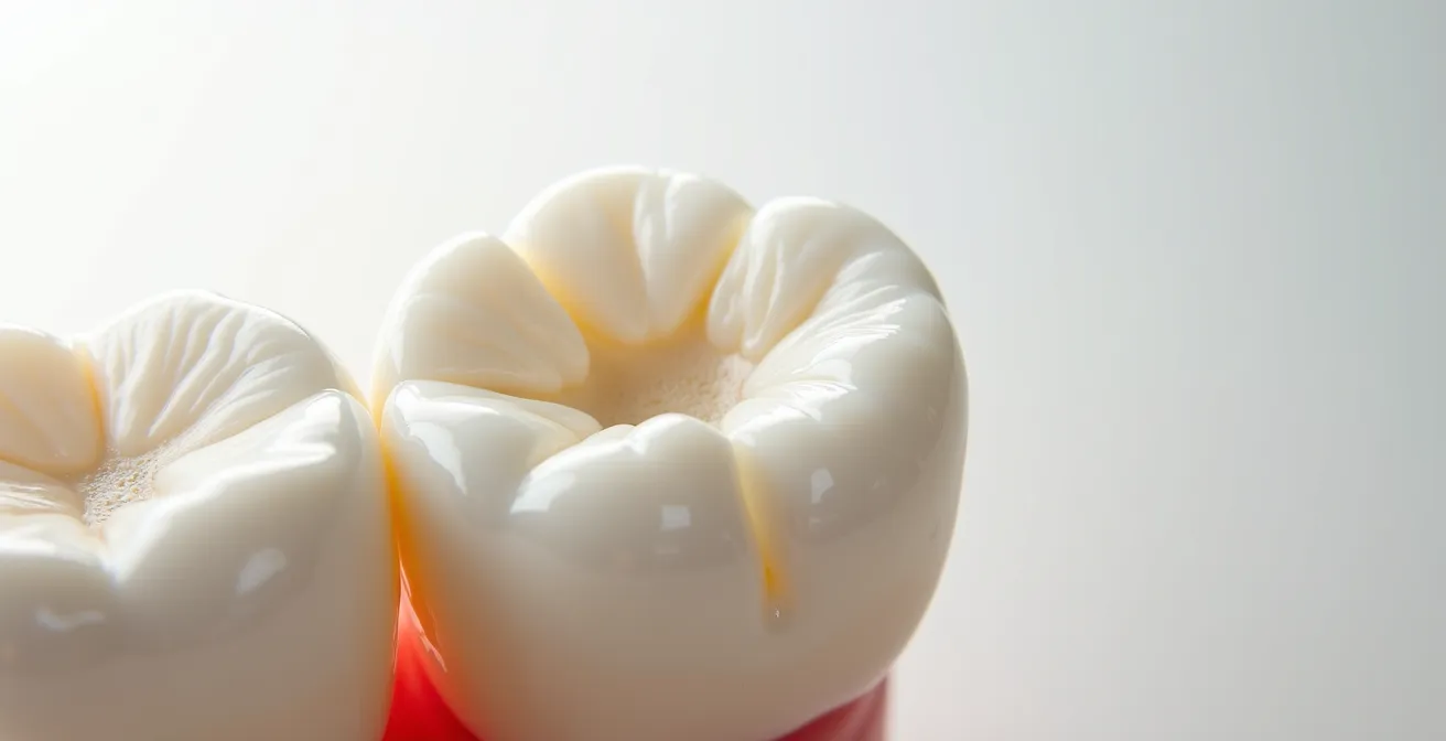
A vague, intermittent ache in a tooth can be one of the most frustrating dental issues. You know something is wrong, but traditional examination methods may not reveal the cause, leaving you and your dentist guessing. This is where high-definition intraoral cameras become an essential diagnostic tool. They act like a dental detective, magnifying the tooth’s surface up to 100 times, bringing microscopic problems into sharp focus.
Often, the source of mystery pain is a hairline fracture, a tiny crack in the enamel that is invisible to the naked eye. When you chew, this crack can flex, irritating the nerve inside the tooth and causing a sharp, fleeting pain. Without visual evidence, this can be misdiagnosed or dismissed. An intraoral camera, however, makes these fractures undeniable. As some of the most advanced digital dental scanning technology in Canada demonstrates, these cameras provide a precise look at hard-to-see places, helping dentists detect the earliest signs of trouble.
The same principle applies to incipient decay (the very first stage of a cavity) or failing restorations. The powerful illumination and magnification can reveal compromised margins on a filling or a spot of demineralization that a traditional dental pick might miss. By making the invisible visible, the camera provides a definitive answer, ending the diagnostic guesswork and allowing for targeted, effective treatment. It transforms your vague symptom into a concrete, solvable problem on the screen in front of you.
How a Photo Can Get Your Denied Dental Claim Approved?
One of the most disheartening experiences for a patient is having a necessary dental treatment denied by their insurance provider. Insurers often operate on a system of codes and written descriptions, which can fail to convey the urgency or necessity of a procedure. A claim for a crown, for instance, might be denied because the written report doesn’t adequately justify why a simple filling won’t suffice. This is where a picture is truly worth a thousand words—and potentially hundreds of dollars.
Intraoral photos provide objective, irrefutable evidence that an insurer cannot easily dismiss. A clear image showing a large, fractured filling or a deep crack running through a tooth immediately demonstrates the structural weakness that necessitates a crown. It removes all ambiguity and proves medical necessity in a way that text alone never could. As the Centre Dentaire de Haute Technologie du Québec in Montreal notes, this visual evidence is a powerful tool in your corner.
The images from the intraoral camera can be stored in the patient’s electronic dental records. We can then refer to them later on or forward them to your insurance company, if required for the approval of your claims.
– Centre Dentaire de Haute Technologie du Québec, Montreal Dental Technology Center
By including these high-resolution images with your pre-determination or claim appeal, your dentist provides a clear, clinical justification for the recommended treatment. This simple step can dramatically increase the likelihood of approval, ensuring you get the care you need without shouldering the full financial burden. It shifts the conversation with your insurer from one of abstract codes to one of concrete, visual facts.
Time-Lapse Dentistry: Monitoring Wear Patterns Year Over Year
Your mouth is a dynamic environment, constantly changing due to factors like diet, stress (leading to grinding or clenching), and natural aging. One of the most significant challenges in dentistry is tracking these slow, subtle changes over time. Are your teeth wearing down faster than they should be? Is that small chip from last year getting worse? Answering these questions used to rely on memory and imprecise plaster models. Today, digital photography enables “time-lapse dentistry.”
By taking a consistent set of intraoral photos at every check-up, your dentist creates a permanent digital baseline of your oral health. This visual archive is invaluable for long-term monitoring. For a patient who grinds their teeth (bruxism), comparing images from year to year can vividly show the progressive flattening of molars or the appearance of new micro-fractures. This visual evidence is far more compelling than simply being told “your teeth are wearing down” and can be the critical motivator for accepting a protective night guard.
This paragraph introduces the intricate details of tooth wear. The illustration below highlights the kind of micro-textures that become visible under magnification, which are tracked over time.
As Montreal dental clinics have found, this method is superior for tracking the stability of restorations, monitoring gum recession, or watching a suspicious area that doesn’t yet require intervention. Having a clear “before” picture provides an objective reference point. Many Montreal dental clinics report that digital archiving enables the comparison of conditions over time, ensuring that small issues are addressed before they become major problems. This longitudinal view transforms dentistry from a reactive practice to a truly proactive and personalized one.
Seeing the Plaque: How Cameras Motivate Better Brushing?
Every dental patient has heard the lecture: “You need to brush and floss more effectively.” While well-intentioned, this verbal advice often lacks impact because the problem—plaque and tartar buildup—is abstract and largely invisible to the patient. An intraoral camera completely changes this dynamic by making the invisible visible and, more importantly, personal.
When a hygienist or dentist uses the camera to show you a magnified view of the fuzzy, yellowish plaque clinging to your gum line or the hard, chalky tartar between your lower front teeth, the need for better hygiene becomes instantly and undeniably clear. The problem is no longer a theoretical concept; it’s right there on the screen, on *your* teeth. This visual confrontation is a powerful psychological motivator. It creates a direct link between the action (or lack of action) of your home care routine and the tangible result in your mouth.
This educational aspect is one of the most significant benefits of the technology. Studies and clinical experience confirm the impact of this visual feedback. In fact, an analysis of in-clinic tools confirms that dental technology studies demonstrate that allowing patients to see their own cavities, plaque, or cracked fillings helps them better understand why a treatment or a change in habit is truly needed. It’s a “show, don’t tell” approach that fosters a sense of ownership over your oral health.
Suddenly, the hygienist’s instructions to angle the brush differently or to wrap the floss in a ‘C’ shape make perfect sense, because you’ve seen the exact areas you’ve been missing. This clear, visual feedback loop is far more effective at creating lasting behavioral change than any lecture could ever be.
Why You Should Run From a Dentist Who Won’t Show You the Photos?
In the modern era of dentistry, transparency isn’t a bonus; it’s the standard of care. An intraoral camera is not just a diagnostic tool but also a cornerstone of patient-provider communication and trust. If a dentist diagnoses a problem, especially one that is asymptomatic and will require an invasive and costly procedure, a refusal to provide visual evidence should be considered a major red flag.
A dentist who is confident in their diagnosis should be eager to show you what they see. Sharing the camera feed is an act of transparency that says, “I want you to be a fully informed partner in this decision.” It respects your autonomy and your right to understand your own body. A reluctance to do so raises uncomfortable questions. Is the problem not as severe as described? Does the evidence not fully support the recommended treatment? Or does the practice simply not prioritize patient education and shared decision-making?
As a patient in Montreal, you have the right to this information. A practice that has invested in this technology but doesn’t use it for patient education is doing you a disservice. A practice that hasn’t invested in it at all may not be keeping up with the modern standard of care. Your role is not to be a passive recipient of care but an active participant. Being empowered starts with asking the right questions.
Your Checklist: Questions to Ask Your Montreal Dentist About Visuals
- Can you show me that issue on the intraoral camera so I can see what you’re seeing?
- Could we take a photo of this area so I can better understand the problem and the proposed solution?
- Will you save these images to my digital file so we can compare them at my next appointment?
- If I proceed with treatment, can you use the camera to show me the before-and-after results?
- How do you use visual technology in this practice to track my oral health over the long term?
Is a 3D Dental Scan Necessary for Routine Implant Surgery?
While intraoral cameras excel at visualizing the surface of teeth, dental implant surgery requires a deep understanding of the structures below the gumline: the jawbone, nerves, and sinuses. For this, another type of imaging is often the standard of care: Cone Beam Computed Tomography (CBCT), or 3D dental scanning. While a traditional 2D X-ray can offer a flat overview, it’s like trying to navigate a city with a simple paper map.
A 3D scan, in contrast, is like having a complete GPS model. It provides a detailed, three-dimensional view of your entire jaw structure. This is particularly crucial for implant placement. A dentist can virtually place the implant on the 3D model before the surgery even begins, ensuring optimal positioning to avoid critical structures like the mandibular nerve or the sinus cavity. It also allows for a precise assessment of bone density, confirming that there is enough healthy bone to support the implant securely.

While it represents a higher upfront cost, a 3D scan dramatically reduces the risk of surgical complications and improves the long-term success rate of the implant. For a procedure as significant as implant surgery, the precision afforded by 3D imaging is not a luxury—it’s a critical component of safe and predictable treatment planning. The difference in diagnostic capability and safety is stark, as this comparison of technologies used in Montreal clinics shows.
| Aspect | 3D i-CAT® Scanning | Traditional X-rays |
|---|---|---|
| View of Jawbone | Complete 3D view | 2D limited view |
| Implant Placement Precision | Optimal positioning determined | Estimation required |
| Nerve Location | Clearly visible in 3D | Difficult to assess |
| Bone Density Assessment | Comprehensive evaluation | Limited information |
| Cost in Montreal | $350-500 CAD | $50-150 CAD |
| Treatment Planning | Precise surgical guide possible | Manual estimation |
Why Do 90% of Adult Cavities Start Between the Teeth?
It’s a frustratingly common dental statistic: the vast majority of cavities in adults form in the interproximal spaces—the tight contact areas between two teeth. The reason is simple access. These areas are shielded from the cleansing action of your tongue and saliva, and toothbrush bristles often can’t reach them effectively. This creates a perfect, undisturbed haven for plaque bacteria to thrive, metabolize sugars, and produce acid that erodes tooth enamel.
Making matters worse, these cavities are also the hardest to detect during a routine visual exam. A dentist can’t see through teeth, so they must rely on bitewing X-rays or the tactile feedback of a dental explorer. However, early-stage interproximal decay might not show up clearly on an X-ray, and a probe can’t fit between tight contacts. This is yet another area where visual technology provides a breakthrough.
These small, high-resolution cameras provide detailed images of the inside of the mouth, offering a level of precision and clarity previously unattainable with traditional dental tools.
– Chestnut Dental, Advanced Dental Care Technology Report
A slender intraoral camera can sometimes be angled to peer into these hidden spaces, revealing the tell-tale chalky white spots of early demineralization or the dark shadow of decay that signals a cavity is forming. As dental imaging research confirms that high-definition cameras reveal details missed by the naked eye, their use is critical for these difficult-to-reach areas. By catching these cavities early, dentists can often recommend more conservative treatments, such as a small resin infiltration or a remineralization protocol, rather than waiting until the decay is large enough to require a traditional drill-and-fill procedure.
Key Takeaways
- Verifiable Proof: Intraoral cameras provide undeniable visual evidence for diagnoses, bridging the trust gap for issues you can’t see or feel.
- Insurance Ally: A clear photo is a powerful tool to prove medical necessity to insurance companies, helping to secure approval for required treatments.
- Long-Term Tracking: Archiving images creates a “time-lapse” of your oral health, allowing for proactive monitoring of wear, cracks, and gum health.
How High-Definition Zoom Finds the Source of Your Mystery Pain?
We began by exploring how high-definition zoom acts as a diagnostic detective, uncovering the hidden sources of mystery pain. But the true significance of this technology isn’t just in *finding* the problem; it’s in how it redefines the patient’s role in the solution. When a dentist shows you the hairline fracture causing your pain, they are doing more than just making a diagnosis. They are inviting you into a transparent process.
This act of sharing visual information is the antidote to the suspicion and uncertainty that many patients feel. It transforms the professional relationship from a paternalistic model (“I’m the expert, do as I say”) to a collaborative one (“Here is what I see, let’s decide on the best course of action together”). This shift is the most profound change that visual technology has brought to dentistry.
Your trust is no longer expected; it is earned through verifiable evidence. Seeing the problem for yourself empowers you to ask more intelligent questions, to better understand the value of the proposed treatment, and to feel confident in your healthcare decisions. It ensures that you are not just a subject in the dental chair, but an active, respected partner in the preservation of your own health. This is the new standard of care you should expect and demand from any modern dental practice in Montreal.
For your next appointment, be prepared to be an active participant. Insist on seeing what your dentist sees and choose a practice that champions this standard of diagnostic transparency and patient partnership.